CLASS: SS 2
SUBJECT: BIOLOGY PERIOD(S) 3:
DURATION: 45 MINUTES
TOPIC: COMPONENTS OF THE MAMMALIAN SKELETON
SUBTOPIC 1: AXIAL SKELETON
SUBTOPIC 2: APPENDICULAR SKELETON
PREVIOUS KNOWLEDGE: STUDENTS AND FAMILIAR WITH THE
TOPIC
OBJECTIVES: At The End Of The Period(s), students should be able to:
Period 1: Describe the components of the axial skeleton. Describe the composition of a typical vertebrate.
Period 2: Name and explain the structures and functions of both the pelvic and pectoral girdle
Period 3: Explain the function of the pentadactyl limbs in man and draw the structures of the forelimbs and hindlimbs.
REFERENCES:
Essential Biology for Senior Secondary schools by M.S Michaels pg 81-91,
Modern Biology for senior secondary school pg 266-269
CONTENT
The skeleton of vertebrates are built on the same basic plan namely.
- The axial skeleton: the skull, vertebral column (backbone) sternum (breast bone) and the ribs makes up the axial skeleton.
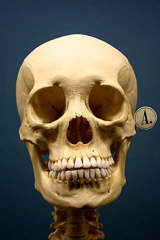
SKULL: The mammalian skull consist consists of several flat bones joined together by sutures (joints) to form the cranium (brain box) which consists of the upper jay (maxilla) and cavities for the main sense organ(eyes, ears and nose) and the lower jaw (mandible) which contains the teeth (alongside the maxilla) and hinges with the rest of the skull in such away that it can move up and down. The functions of the skull include
- it protects the vital sense organs and the brain
- it gives shape to the head
iii. it bears the teeth used for chewing and grinding food
The base of the skull pivots on the top vertebra of the vertebral column which enables the nodding and rotation of our heads.
- Vertebral column: serves as the central supporting structure of the skeleton. it forms the backbone and protects the spinal cord. The human vertebral column consists of 33 vertbera (46vertebra in rabbits) which form a flexible column with a central canal for the spinal cord. Discs of fibocartilage separates the vertebrate which are held together by strong ligaments called intervertebral discs which allow the vertebrae to more slightly, either forwards a backbone or sideways. Openings between adjacent vertebra allows the entry and exit of spinal nerves. A typical vertebra consists of the following.
- A neutral spine which projects upwards dorsally for muscle attachment.
- A neural canal for the passage of the spinal cord.
- the transverse processes which project from the sides of each vertebra for muscles and ligaments attachment
- The centrum which is a solid piece of bone below the neural canal
- Facets (smooth surfaces) at the front and back which fit into the adjacent vertebrate
|
|
Types of vertebrate |
Body of region |
Man |
Rabbit |
Rat |
Characteristic features of each type of vertebrate |
|
1 |
Cervical vertebra |
Neck |
7 |
7 |
7 |
The first cervical vertebra (atlas) allows the head to nod and the second cervical vertebra (axis) allows turning of the head. The cervical vertebrate contain a pair of openings (vertebratenial canals) for the neck blood vessels. The transverse processes are flattened out and are called cervical ribs. |
|
2. |
Thoracic vertebrae |
Chest |
12 |
12 |
13 |
It aids ribs attachment and assists in breathing. Muscle of the shoulder end back and attached to the neural spines. It has a large central, large neural canal, big neural |
|
3. |
Lumbar vertebra |
Lion |
5 |
7 |
6 |
It provides (has processes) attachment for abdominal muscles and bear considerable weight of the body lumbar vertebrate has a large centrum and a neural spinethat projects upwards and forwards. |
|
4 |
Sacral vertebra |
Hip |
5 |
3-4 |
4 |
Sacral vertebrae are joined to the pelvic girdles toprovide support, rigidity and strength. Sacral vertebrae fuse to form a rigid structure, the sacrum. The sacral canalis narrow and the neural spine is reduced. |
|
5 |
Caudal vertebra |
Tail |
4 |
16 |
27-30 |
The caudal vertebrae are found to form a solid mass of bone. It support the tail and provides attachment for the tail muscle. |
|
|
Total |
|
33 |
45-46 |
57-60 |
|
RIBS AND STERNUM – The sternum and 12 pairs of ribs form the rib cage. The sternum is a single bone in man but in rabbits, it consists of 7 small bones. The ribs articulate with the thoracic vertebrae at the back of curve to the front to attach by means of cartilage to the sternum. In man, just 10 out of the 12 rib. (First 7 are true ribs because they are attached directly, 8,9, 10 ribs are false ribs because they have a common connection toteh sternum. Pairs are attached to the sternum. The remaining 2 paris remains free and are referred to as floating ribs. Rabbits have 3 floating ribs.
Human skull

Lumbar vertebra (posterior view)

2 The appendicular skeleton is made up of the limb girdles (pectoral and pelvic girdles) and the limbs (forelimbs and hindlimbs)
Pectoral girdle: it holds the upper limbs (arms) to the axial skeleton. It consists mainly of 4 separate bones namely two large flat triangular sholder blades or scapulae (sing scapular) at the back and two small slender collar bones or clavicles in front. The scapulare are attached to the spinal columb by muscles. A hollow or depression, glenoid cavity, is found at the apex of the scapula into which the ehad of the humerus (upper arm bone) fits to and from the shoulder joint. Above the glanoid cavity is a small hook shaped bone called the coravcoid bone. The clavicles are attached to a scapula at one end and to the sternum at the other end. The pectoral girdle is not rigid, enabling the arms and shoulders to move fairly freely thereby providing firm support for the forelimbs
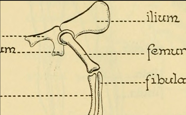
Pelvic Girdle: It is found around the waist in man. The pelvic girdle(hips) consists of two bones, the right and left pelvis joined to the sacrum (dorsally) at the back and held together by fibro-cartilage at the facets in front to form a complete rigid girdle. The kind of fusion of the two bones is called pubis symphysis. Each bone is made up of three bones (ilium, ischium and pubis) which are fused together.
The ilium which is the longest and largest of the three bones are at the top. Ischium and pubis (the fused bones) enclosed an opening called obturator foramen through which nerves blood vessels and muscles pass. A deep cavity or hollow at the outer edge of each pelvis (where the 3 bones meet) the acetabulum, into which the head of the thigh bone (femur) fits to form the hip joint (a ball and socket joint) The pelvic girdle receive the weight of the upper body and pass it on to the legs (if standing) or to the surface on which you are sitting. The movements of the hips and legs are restricted by the rigid structure of the girdle.
THE LIMBS: the limbs of most mammals are built on the same basic pentadactyl limb plan (5-digit plan) it consists of a long bone, followed by a pair of long bones placed side by side , a set of nine small bones in three rows, five thin long bones and finally five digits. Each digit is madeup of a few small bones.
The forelimbs (arm) consists of the upper arm, lower arm, wrist and hand. The upper arm bone (humerus) which is a long bone has a rounded head fits and articulates with the glenoid cavity of the scapular in the shoulder joint. The humerus articulates with the radius and ulna (the two bones of the fore/lower arm) to form the elbow joint. The radius is a long, slightly curved bone which lies in front of the ulna. The ulna is longer than the radius and has a cavity (sigmoid cavity) into which the trochlea of the humerus fits. The ulna also project backwards to form a projection called olecranon process. The bones of the wrist (9small bones arranged in 3 rows)/carpals followed by the five digit bones (metacarpals) called fingers/phalanges in man. Each digit has three phalanges except thumb which has 2. The ulna and radius can partly rotate around each other so that the hand can be held pal upwards or downwards.

Diagram of the pentadactyl limb (Right) the left front limb of rabbit.
The hindlimb (leg) consists of the thigh, shank, ankle and foot. The thigh bone (femur) is the largest and strongest bone in the body. The femur head at the proximal end fits into the acetabulum of the pelvic girdle to form a hip joint. Three projections (trochanter) very close to the femur head are used for muscle attachment. At the distal end of the femur, the condyles (rounded nobs) articulate with the tibia bone and fit into two grooves in the tibia. The shank has 2 bones, tibia and fibula (fused in rabbit tibio-fabula). The tibia is longer and larger than the fibula (the fibula lies outside the tibia). The knee joint is found at the junction of the femur and the tibia. The knee cap (patella) is present at the front of the knee joint. The ankle is made up of six bones called tarsals. The inner tarsals project backward to form the heel bone. Most mammals including man have five metatarasals (digits). The bones of the foot are not as flexible (dexterous) as those of the hand but form an arch which enables the foot to withstand tremendous forces.
INSTRUCTIONAL MATERIALS: A replica of the mammalian skeleton. Pictures and videos showing the skeleton
TEACHING METHODS PROCEDURES
Step I: Teacher revises the previous topic and introduce the new topic
Step II: Teacher explains the components of the mammalian skeleton – the axial and appendicular skeleton
Step III: Teacher explains using the replica of the mammalian skeleton
Step IV: Teacher allows students to ask questions and write notes under content on the board.
ACTIVITIES
Period 1: Students discuss the components of the mammalian skeleton.
Period 2: Students with the help of the teacher discuss the axial skeleton
Period 3: Students discuss the appendicular skeleton
EVALUATION
Period 1: What is a skeleton?
Period 2: What are the components of the mammalian skeleton?
Period 3: The forelimb consists of how many parts?
SUMMARY: Teacher summarizes the topic, marks the students notes and make corrections where necessary
ASSIGNMENT:
Period 1: State five differences between bone and cartilage
Period 2: Briefly discuss the skeletal system of mammals and give examples
Period 3: Name the major bones of the appendicular skeleton and describe one of them in details.