Groups
-
david-manglideposted in News & Trends • read more
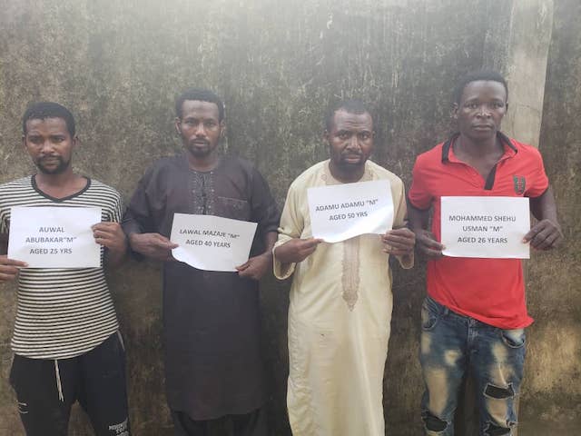
The Nigerian Police have unveiled the faces of four suspects said to be Funke Olakunrin’s killers.
Olakunrin, daughter of Pa Reuben Fasoranti, was killed on 12 July 2019, at Kajola on Ondo-Ore Road by gunmen suspected to be Fulani marauders.
After some painstaking investigation, IGP Mohammed Adamu announced the arrest of the suspects on Thursday.
They are: Lawal Mazaje,40, from Felele, Kogi and Adamu adamu, 50, from Jada in Adamawa.
The others are Mohammed Shehu Usman, 26, from Illela Sokoto and Auwal Abubakar, 25, from Shinkafi, Zamfara state.
However, the IGP also announced that four other suspects, including the ringleader named Tambaya, are at large.
The Police have declared wanted and have given out some description.
According to the police, Tambaya speaks Hausa, Fulfulde and Pidgin English.
“He is fair in complexion and in his late 20s – between the age of 27 and 30.
“His last known address is Isanlu, Kogi State.
“He has visible scar from stitches on his forehead down to his nose and mouth”.
-
david-manglideposted in News & Trends • read more
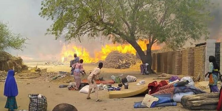
No fewer than 14 people have been burnt to death at an Internally Displaced Persons, IDP, Camp in Borno State.
The blaze broke out mid-morning in a camp holding some 70,000 people in the town of Gamboru, close to the border with Cameroon, the head of a government-backed militia told AFP.
“We recovered 14 burnt bodies from the gutted shelters and another 15 who sustained varying degrees of burns from the fire,” militia leader Umar Kachalla said.
More than 200 shelters were destroyed, rendering 1,250 people homeless, said a rescue official, who asked not to be named.
“The fire killed 14 people, six from the same family, and left 15 with injuries, seven of them critically,” said the official.
It was not clear what caused the fire but residents blamed it on arson after a suspect was arrested with a lighter in his pocket, another militia member Shehu Mada said.
The decade-long Boko Haram conflict has killed 36,000 people and forced 1.8 million from their homes in northeast Nigeria.
Hundreds of thousands of people have been left living in often squalid and overcrowded camps where they rely on food aid from international charities, AFP reports.
-
david-manglideposted in News & Trends • read more
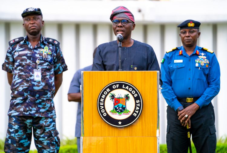
Lagos State Governor, Babajide Sanwo-Olu on Thursday disclosed that five more Coronavirus patients have been discharged from isolation centre in the state.
The governor said those discharged included three female and two male.
“Dear Lagosians, Today, 5 more patients; 3 females and 2 males, have been discharged from the Mainland Infectious Diseases Hospital to reunite with the society.
“They were discharged having recovered fully and tested negative twice consecutively to #COVID19,” he said.
According to Sanwo-Olu, this brought to 90, the number of patients successfully managed and discharged from the State’s isolation facilities.
“I appeal to residents to stay at home, practice Social Distancing and observe the highest possible personal and hand hygiene,” he said.
-
david-manglideposted in News & Trends • read more
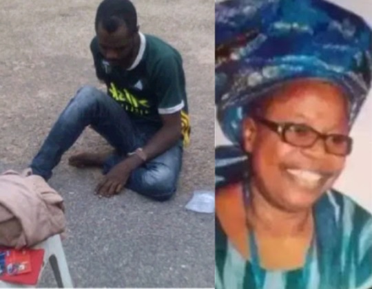
A 34-year-old murder suspect, Abegunde Olaniyi has confessed to killing an evangelist with the Christ Apostolic Church, Oke Imole, Ibadan, Oyo State, Mrs Grace Ajibola after being paraded on Tuesday by the Oyo State Police Command..
Olaniyi, who was described as an errand boy to the evangelist, said he first hit her with a wooden object and then strangulated her, and thereafter carted away her Tecno mobile phone and three Automated Teller Machine (ATM) cards.
He said he had discovered Mrs Ajibola had a sum of about N2 million in her bank account after she sent him to withdraw money for her via the ATM at a new generation bank at Apata, Ibadan.
“I decided to murder her and take possession of the money,” Olaniyi said.
He said he had already withdrawn the sum of N120,000 from the victim’s bank account when he was arrested.
The Oyo State Commissioner of Police, Sina Olukolu, told journalists yesterday at the Command Headquarters, Ibadan, that Olaniyi killed Mrs Ajibola on March 17, 2020 at about 11am at her residence at the Oluyole area of Ibadan.
He said the police recovered one Tecno mobile phone and two ATM cards belonging to Mrs Ajibola and the wooden object that the suspect used in hitting her.
The police boss added that the suspect was arrested in his hideout in Akure, Ondo State.
Olaniyi told journalists he didn’t know what came over him on that fateful day that made him kill the evangelist.
-
david-manglideposted in News & Trends • read more

Fuji Icon, Wasiu Ayinde popularly known as KWAM 1 has reacted to rumours that he has been having an extramarital affair with the youngest Queen of Alaafin of Oyo, Queen Badirat.
KWAM1, who was recently given a title by the Alaafin of Oyo, Oba Lamidi Adeyemi, gave his reaction via short music video posted on his Instagram page.
According to him, the rumour was cooked up by those working day and night to see his downfall.
He accused the rumour mongers of trying to tarnish his image since he became the Mayegun.
Kwam1 further charged his fans not to react or contend with the false claims, as such won’t influence his status or his music profession that had spread over decades.
-
david-manglideposted in News & Trends • read more
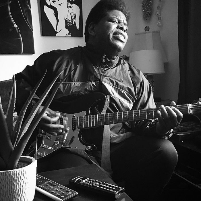
Menegian, son of late activist, Ken Saro-Wiwa has died of Coronavirus in the United Kingdom.
This was disclosed by his sister, Noo Saro-Wiwa on her Facebook Page on Thursday.
According to Noo, his bother had Coronavirus, combined with underlying health conditions, on Monday, which led to his death.
“We said goodbye to my brother, Gian; on Monday, he had COVID-19 combined with underlying health conditions. Gian was the smartest and most talented out of all of us.
“A champion sprinter at school, a poet, an artist, budding engineer, a self-taught guitarist and pianist. But mental health issues limited his life from age 16 onward.
“I took this photo of him a few months ago before he was hospitalised. We were singing Hysteria by Def Leppard, a song we both love. Although the side-effects of medication altered his athletic physique, this photo still captures Gian’s essence: a kind and beautiful soul, always wanting the best for his family, always praying for our future success while being eternally optimistic about his own.
“And, of course, always loving his music. He leaves a wonderful son, Louis,” she said.
Menegian Saro-Wiwa was born in 1970.
-
david-manglideposted in News & Trends • read more
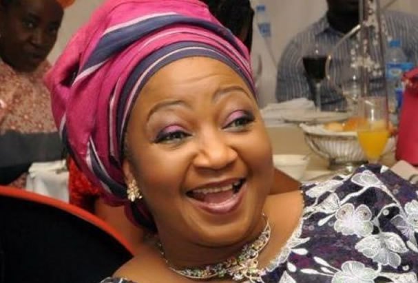
Four men suspected to have murdered Mrs Funke Olakunrin, daughter of Pa Reuben Fasoranti have been arrested.IGP Mohammed Adamu announced the arrests of the four suspects on Thursday.
But he said four other suspects, including the ringleader named Tambaya are on the run.
Adamu has declared Tambaya a wanted man.
Those arrested are: Lawal Mazaje,40, from Felele, Kogi and Adamu adamu, 50, from Jada in Adamawa.
The other two are Mohammed Shehu Usman, 26, from Illela Sokoto and Auwal Abubakar, 25, from Shinkafi, Zamfara state.
Funke Olakunrin was killed at Kajola on Ondo-Ore Road on 12 July 2019.
The murder, suspected to be by Fulani marauders triggered national uproar.
The police in a statement explained how the suspects were nabbed by a police crack team led by CP Fimihan Adeoye.
JUST IN: Police arrest Funke Olakunrin’s killers, 4 other suspects still on the run
Thursday, April 16, 2020 4:03 pm | Daily News Headlines | 0 Comment(s)
Late Mrs. Funke Olakunrin: four suspects arrested
Promoted Content
Mgid
Earn $1300 Per Day With Amazon Shares, $200 Minimum Investment
investstocks
While Stocks Are Falling, Nigerians Are Getting Rich Doing This
IQ Option
I Just Invested $200 In Amazon Shares & Now I Earn $967 A Day
investstocks
Top 10 Worst Jobs In The World That Someone Has To Do
Red Dragons MediaFour men suspected to have murdered Mrs Funke Olakunrin, daughter of Pa Reuben Fasoranti have been arrested.
IGP Mohammed Adamu announced the arrests of the four suspects on Thursday.
But he said four other suspects, including the ringleader named Tambaya are on the run.
Adamu has declared Tambaya a wanted man.
Those arrested are: Lawal Mazaje,40, from Felele, Kogi and Adamu adamu, 50, from Jada in Adamawa.
The other two are Mohammed Shehu Usman, 26, from Illela Sokoto and Auwal Abubakar, 25, from Shinkafi, Zamfara state.
Funke Olakunrin was killed at Kajola on Ondo-Ore Road on 12 July 2019.
The murder, suspected to be by Fulani marauders triggered national uproar.
The police in a statement explained how the suspects were nabbed by a police crack team led by CP Fimihan Adeoye.
“After months of relentless efforts to apprehend the killers, the Police Team, on 4th March, 2020 during a follow-up action on a case of a high-profile armed robbery and kidnap for ransom that occurred in Ogun State, arrested one Auwal Abubakar ‘m’ 25yrs, an accessory after the fact of the crime, along Sagamu-Ore expressway in Ondo State.
“The arrest of Auwal Abubakar led to the arrest of two other members of the gang, Mohammed Shehu Usman and Lawal Mazaje in Benin, Edo State from whom cache of ammunition was recovered and one other Adamu Adamu in Akure, Ondo State.
“Having established sufficient physical and forensic evidence linking the suspects to the killing of Mrs Funke Olakunrin, the investigators, determined to clear all doubts relating to their findings.
“On 8th April, 2020, they conducted an Identification Parade at the Federal SARS Headquarters, Lagos which led to the positive and physical identification of three (3) suspects, Adamu Adamu, Lawal Mazaje and Mohammed Shehu Usman by a survivor of the earlier crime.
“The survivor gave a clear description of the roles each of the identified suspects played in the killing.
“At this point, the suspects capitulated and voluntarily offered a no-holds-barred confession on how Mrs Funke Olakunrin was killed.
“Investigations so far reveal that the operation that led to the killing was carried out by eight fully armed kidnap/robbery suspects led by one Tambaya (other name unknown) who is currently at large.
“While four of the suspects are in custody, effort is being intensified to arrest the four others still on the run.
“The 8-man gang has its operational base and membership spread in the south-western part of the country and Edo State.
“Investigations have also revealed that they are responsible for series of high-profile armed robbery and kidnap operations in the region.
“They also attack, vandalize and steal components of critical national infrastructures such as electrical and telecommunications installations.
“Consequently, the Inspector General of Police hereby declares the principal suspect, Tambaya (other names unknown) wanted for his involvement in the death of Mrs Funke Olakunrin.
“Tambaya, a Nigerian, speaks Hausa, Fulfulde and Pidgin English.
“He is fair in complexion and in his late 20s – between the age of 27 and 30.
“His last known address is Isanlu, Kogi State. He has visible scar from stitches on his forehead down to his nose and mouth”.
-
david-manglideposted in SS 3 • read more
CLASS: SS3
SUBJECT: BIOLOGY PERIOD(S) 3
TOPIC: MECHANISM OF TRANSPORT IN HIGHER PLANTS
SUBTOPIC 1: TRANSLOCATION AND TRANSPIRATION
SUBTOPIC 2: WATER ABSORPTION AND TRANSPORT
SUBTOPIC 3: DIFFERENCES IN PLANT AND ANIMAL TRANSPORT
PREVIOUS KNOWLEDGE: Students are familiar with the topic
OBJECTIVES: At the end of the period
- Students should be able to:
PERIOD 1:
- Discuss the mechanism of transportation in plants
- Describe the tissues involved in the transportation of material
PERIOD 2:
- Explain how manufactured food, water, mineral salts, and other materials are transported in plants.
PERIOD 3:
- List the differences between transport in plants and animals.
- Demonstrate experimentally the flow of materials in plants
REFERENCES:
Essential Biology for Senior Secondary Schools by M.C Micheals pg 292 – 300
Modern Biology for senior secondary schools pg 312 – 317
CONTENT
In a simple plant (algae), materials enter or leave the body cells by diffusion (gases) manufactured food and waste products) and water enters by osmosis. In higher land plants, vascular tissues (special conducting tissues) carry out transport with latex tubes to assist. The major materials transported in plants are gases (CO2 and O2), water, mineral salts, manufactured food, nitrogenous waste products, hormones and pigments. Plant saps, cell sap and cytoplasm are the main transport media. Gases are mainly absorbed through the stomata (in the leaves) and lenticels (in the stem).
Water and mineral salts are absorbed through the root system, water, mineral salts and soluble food are transported in the vascular tissues of the plant. The vascular tissue are made up of a network of long tubes called vascular bundles. A vascular bundle consists of the xylem and phloem tissues. The cambium exists between the xylem and phloem tissues in the roots and stems of dicotyledonous plants.
- Cambium – are made up of narrow living cells with thin walls and dense cytoplasm. Cambium produce secondary xylem and phloem by dividing and multiplying. This growth in width (girth) of the stem is called secondary thickening.
- Xylem tissues – consists of dead cells with lignified cell walls. Xylem transport water and dissolved mineral salts from the roots to other plant parts. It also give support and rigidity to plants.
iii. Phloem: Consists of thin walled living cells with dense cytoplasm which have perforated cross walls. Phloem transports a manufactured food from the leaves to other plant parts.
TRANSLOCATION
This is the process by which manufactured food is transported from green leaves of a plant to all the living plant cells especially those in the actively growing regions (root tips, stem tips and buds) and excess food is also stored. Phloem is responsible for this. Glucose, oil, resin, protein, alkaloids and hormones are among the translocated materials.
TRANSPIRATION
Transpiration is the loss of water from the leaf surface of plants by evaporation. The continuous flow of water from the roots to the leaves is called the transpiration stream. Plants loose excess water through the stomata (stomata transpiration), lenticels (lenticular transpiration) and the leaf cuticle (cuticular transpiration). Transpiration is affected by the following factors:
- Temperature – increase in temperature causes increase in transpiration rate
- Wind – increase in wind causes increase in transpiration
iii. Soil water-high level of soil water results in higher absorption and transpiration rate.
- Light – High light intensity results in high photosynthetic rate leads to increase in temperature and therefore, increase in transpiration.
- Humidity: Higher humidity leads to reduce rate of transpiration and vice versa.
- size of stomata pores – when the guard cells become turgid and the stomata open leading to transpiration however when the guard cells are flaccid, the stomata close and transpiration stops.
WATER ABSORPTION BY PLANT ROOTS
The cell sap in the root hairs is more concentrated than the soil water. The cell membrane of the root hairs is selectively permeable therefore, soil water enters the vacuole of the root hairs via osmosis. The extra water causes increase in tugor pressure of the vacuole (reduce osmotic pressure) and forces water out into the cell walls towards the cortex. The cell next to the root hair on the inside has a lower turgor (higher osmotic pressure) hence water will pass into it by osmosis. This process enables water absorbed to get to the xylem vessels.
TRANSPORT OF WATER IN THE XYLEM TISSUE
Water transport by the xylem is due to the following process
- Capillary action: This allows the upward movement of water through the xylem from the roots to the leaves. The xylem vessels form very fine capillary tubes.
- Root pressure and suction pressure – Osmotic pressure difference between the cell sap and soil nutrients concentration create root pressure. The cell sap becomes more concentrated and it tends to draw up the nutrients. Suction pressure occurs during stomata transpiration. This pressure facilitates the movement of water from the soil to the xylem tissues.
iii. Transpiration stream or pull
SIMILARITIES IN TRANSPORT IN ANIMALS AND PLANTS
- A liquid medium is required
- Materials are transported in dissolved form.
- Tabular or cylindrical vessels are necessary
- Diffusion and Osmosis play key roles in transportation
DIFFERENCES BETWEEN PLANTS AND ANIMALS
PLANTS
ANIMALS
1
Media from transportation is cell sap
Blood is the medium of transportation `
2
Root pressure or transpiration generates forces for pull
Heart generates forces for transport of nutrients.
3.
The transport medium is not tissue
Transport medium is made up of cells of different types of tissues.
4.
Materials are transported through different vessels (xylem and phloem)
Materials are transported in the same vessels(blood vessels)
INSTRUCTIONAL MATERIALS – PICTURES AND VIDEOS ON TRANSPORTATION INPLANTS
TEACHING METHODS PROCEDURES
Step 1: Teacher revises the previous topic and introduce the new topic
Step II: Teacher explains how materials get transported in plants.
Step III: Teacher explains absorption of water and mineral salts by the root hairs.
Step IV: Teacher lists the differences between transportation in plants and animals.
Step V: Teacher allows students to ask questions and writes notes under content on the board.
CLASSWORK (ACTIVITIES)
PERIOD 1: Students discuss transportation of materials in higher plants
Period 2: Students discuss the vessels of transportation and how they carry out their functions
Period 3: Students list the differences in plants and animal transport.
EVALUATION
PERIOD I: Explain transport in higher plants
PERIOD 2: State the function of
- Xylem
- Phloem
PERIOD 3: List 3 factors affecting transpiration
SUMMARY/CONCLUSION: Teacher summarizes the topic, marks the notes of the students and make corrections where necessary.
ASSIGNMENT(S)
- Describe an experiment to show the translocation occurs through the phloem tissue.
- Describe an experiment to demonstrate transpiration in plants.
- Describe an experiment to show that water is conducted in the xylem tissue of flowering plants.
- Describe an experiment to demonstrate root pressure and transpiration pull.
-
david-manglideposted in SS 2 • read more
CLASS: SS2
SUBJECT: BIOLOGY PERIOD(S): 3
TOPIC: CIRCULATORY SYSTEM IN MAMMALS
SUBTOPIC 1: Structure of the circulatory system
Subtopic 2: Types of circulation
Subtopic 3: Mechanism of circulation
Previous Knowledge: Students are familiar with the topic
Objectives: at the end of the period(s) students should be able to
PERIOD 1: Describe the structure of the components of the circulatory system
- Describe the types of circulation
PERIOD 2: Compare and contrast the various mechanisms of transportation.
REFERENCES: Essential Biology for Senior Secondary Schools 1-3 MC Michaels pg 285 – 292
Modern Biology for Senior Secondary Schools 1-3 pg 301 – 311
CONTENT
Circulatory system refers to the continous movement or flow of blood round the body involving the heart and the blood vessels. A circulatory system has the following features.
- A circulatory fluid, which is usually blood
- A pumping device or heart to drive the fluid around the body and
- A system of branched tubes or vessels connected to the heart through which the fluid can circulate.
There are 3 major categories of circulatory system in animals namely:
- Open and closed circulatory system
- Single and double circulatory system
- Pulmonary and systemic circulatory system.
Open circulatory system – can be found in some animals like mollusks and insects. The heart pumps blood out into a blood vessel which branches and opens into spaces in the body cavity called haemocoels. The organs and tissues are bathed in blood and exchange of materials occurs between the body cells and the blood. Blood in the spaces eventually flow into the vessels leading to the heart. Valves in the heart and vessels flow in one direction .
Closed circulatory system –
This system can be found in all vertebrates and some invertebrates like annelids. The blood is contained within vessels and is always confined within the cavities of the vessels and heart and never coems in contact with the body cells. Arteries carry blood from the heart and branch and enter various organs and tissues where they divided into smaller vessels (arterioles) which branch again and again to form fine vessels (capillaries). Capillaries are found in between all body cfells and they exchange materials with body cells by diffusion. Capillaries unit to form venuoles which joine together to form vein which carry blood back to the heart. Oxygenated blood is confined to the left part ofhte heart and leaves the heart at high pressure while deoxygenated blood is confined to the right part of the blood and returns to the ehart at low pressure.
Single circulatory system: The Blood passes through the heart only once everytime it makes one complete movement round the body. This system is common in fish which has only two chambered heart (One auricle and one ventricle each) The single circulation is not efficient. The arteries form capillary network at the gills and also at the organs and tissues. Each time this happens, the blood pressure decreases and blood that collects in the veins is at low pressure and move sluggishly. To help blood flow in the veins leading to the heart, valves are present in veins and some veins are replaced by blood spaces.
Double circulatory system
The blood passes through the heart twice every time it makes one complete movement round the body and each time the blood passes through the heart, it goes through a separate pathway. Double circulation gives rise to two pathways namely:
- Pulmonary Circulation – Blood circulates from the heart to the lungs and back to the heart
- Systemic circulation : Blood circulates from the heart to all other parts of the body and back to the heart.
In one complete circulation, blood from any part of the body enters the heart for the first tiem and sent to the lungs for oxygenation. From the lungs, it is brought back to the heart and second time befor it is pumped to all the parts of the body. Frogs exhibit partial double circulation since the oxygenated and deoxygenated blood are not completely separated as in mammals
BLOOD VESSELS
Blood vessels are tubes which transport blood to or from the ehart and there are 3 major types of blood vessels namely:
- Arteries – have thick, muscular, elastic walls which are capable of withstanding high pressure of the blood (Blood is at high pressure of the blood (Blood is at high pressure, flows fast and in spurts) Arteries transports oxygen blood from the heart except that in the pulmonary arteries. Arteries are capable of constricting or circulating and thereby regulate the amount of blood. Valves are absent except for the semi-lunar valves near the heart.
- Veins have fairly thick slightly elastic wall with large lumen. Veins carry deoxygenated blood back to the heart except the pulmonary vein. The blood is at low pressure, flows slowly and smoothly valves are present to prevent backflow of blood. The flow of blood is assisted by the movement of the skeletal muscles.
- Capillaries are tiny blood vessels (One cell thick) which links arteries to the veins. They are found in the junction between arteries and veins around tissues and organs in the body. Capillaries have thin walls which allows easy diffusion of dissolved food substances, waste products and oxygen. Blood pressure is hgihter at arterioles and then at the venules end of the capiilariy network. Therefore blood flows smoothly. The blood is oxygenated at arteriole end and deoxygenated at venule and except that in the lung capillaries.
DIFFERENCES BETWEEN ARTERY AND VEIN
ARTERY
VEIN
1.
Has thick, muscular, elastic walls
Has thin, less muscular, non elastic wall
2.
Carries blood away from the heart
Carries blood to the heart
3.
Carries oxygenated blood except the pulmonary artery
Carries deoxygenated blood except the pulmonary vein
4.
Blood in it is pink or bright red
Blood in it is dark red in colour
5.
It is situated deep in the muscles
It is superficially located
6.
It has a small lumen
Has large lumen
7.
Pressure is high and pulse is readily detectable
Pressure is low and pulise is not readily detectable
8.
Possess no valves except semiluna valves
It has valves
9.
Starts at large vessels and subsequently subdivide
Starts as tiny vessels and subsequently mere into large vessels
THE HEART
The heart is a conical hollow, muscular organ which works continuously pumping blood round the body throughout an organisms lifetime. It is about the size of a clenched fist and weighs about 300grams. The heart is located in the chest (thoracic cavity) just behind the breast bone (sternum ) and between the lungs and it is made up of special muscles (cardiac muscles) and enveloped in a two layered tough protective membranous bag, the pericardium. Pericardial fluid fills the space between the 2 layers.
The pericardial fluid reduce friction caused by the heart’s pumping action. Each pumping action of the heart is called the heartbeat which is caused by the alternate contraction and relaxation of the four heart chambers. Heartbeat occurs in 2 stages namely:
- Diastole: This is the first stage during which the 2 auricles contact creating a high blood pressure. This pressure causes the bicuspid and tricuspid valves to fold downwards into the ventricles, allowing deoxygenated blood to enter the right ventricle from the right auricle and oxygenated blood to enter the left ventricles from the left auricle. Once the ventricles are full, the cuspid valves close up the apertures between the upper and lower chambers of the heart.
- Systole – this is the second stage which involves the contraction of the two ventricles which results in the blood being forced into the two trunks of the main arteries and out of the heart. Deoxygenated blood from the right ventricle passes into the pulmonary artery while oxygenated blood from the left ventricle passes into the aorta.
When at rest, the average human heart contracts and relaxes about 70-72 tones per minute (up to 100 or more during activity, 200 heartbeats per minute in small animals, 12 in big animals like elephant0 and pumps about 14,000 litres of blood a day.
STRUCTURE OF THE HEART
The hearts has 4 chambers: 2 upper this walled atriacle or auricles and 2 lower thick walled ventricles. The right side of the heart is separted form the left by a wall called septurn. Two large veins, the superior (anterior) and inferior(posterior) venae cavae supply deoxygenated blood from parts of the body (except the lungs) into the right auricles. The pulmonary veins supply oxygenated blood into the the left auricle. The right auricle opens into the right ventricle and this is controlled by the tricuspid valve which has 3 flaps which are attached by chordate tendinae (cord like tendons) to the right ventricle walls which allow unidirectional blood flow from the right auricle to the right ventricle. Bicuspid or mitral valve controls the opening of the left auricle into the left ventricle (has 2 flaps) The right ventricle opens into the pulmonary artery, which branches into 2, one to the right lung and this other to the left lung and they carry deoxygenated blood to the lungs. The left ventricle opens into the large aorta (largest artery) which branches to distribute oxygenated blood to the parts of the body except of the longs. Semilunar valves in tehse arteries prevents blood backflow into the ventricles. Blood enters the aotar at a very high pressure (about 105mmHg) and blood entering the pulmonary artery is at a much lower pressure (about 16mmHg) It is because the contraction of the left ventricle must send blood round the much longer systemic circulation therefore the wall of the left ventricle is at least three times thicker than that of the right ventricle.
Examples of major arteries in the body and the organs they supply
Blood vessel (Artery)
Organ supplied
1
Carotid Artery
Head
2.
Pulmonary Artery
Lungs
3
Subclanan artery
Forelimbs
4.
Hepatic artery
Liver
5
Mesentric artery
Stomach and intestine
6
Renal artery
Kidney
7
Gonadial Artery
Gonads
8.
Intercoastal arteries
Wall of thorax
9.
Iliac artery
Hind limbs
INSTRUCTIONAL MATERIALS: Pictures and videos
TEACHING METHODS PROCEDURES
Step 1: Teacher revises the previous topic and introduce the new topic
Step II: Teacher defines circulatory system and mentions the categories of the circulatory system
Step III: Teacher explains the function of each blood vessel in detail and mention the differences between an artery and a vein
Step IV: Teacher describes the structure and function and the mammalian heart.
Step V: Teacher allows students to ask questions and write note under content on the board.
CLASSROOM (ACTIVITIES)
PERIOD 1: Students discuss circulatory system in man.
PERIOD 2: Students discuss the types of circulatory system and blood vessels
PERIOD 3: Students discuss the structure and function of the heart.
EVALUATION ;
Period 1: Mention the components of the mammalianblood
Period 2: List 4 differences between artery and vein
Period 3: How many times does a man’s heart beat in a minute
SUMMARY/CONCLUSION: Teacher summarizes the topic, marks the students notes and makes corrections where necessary.
ASSIGNMENT(S)
Period 1: Describe three ways by which the blood protects the body against diseases causing organisms
Period 2: Make a large labeled diagram of the outline of the blood circulatory system in mammals.
Period 3: Draw and label the mammalian heart.
-
david-manglideposted in SS 2 • read more
CLASS: SS2
SUBJECT: BIOLOGY PERIOD(S): 3
TOPIC: TRANSPORT SYSTEM
SUBTOPIC 1: MATERIALS FOR TRANSPORTATION
SUBTOPIC 2: MEDIA OF TRANSPORTATION
SUBTOPIC 3: COMPOSITION AND FUNCTIONS OF BLOOD AND LYMPH
PREVIOUS KNOWELDGE: Students are familiar with the topic
OBJECTIVES: At the end of the period(s) students should be able to
Period 1:
- Explain the necessity of a transport system in complex organisms
- Explain the in adequacy of diffusion alone as a transport system for complex organisms
Period 2:
- Identify the source of materials for transportation
- Identify where there materials are transported to:
Period 3: Discuss the different types of transportation media such as cytoplasm, cell sap, lymph and blood.
References :
Modern Biology for Senior Secondary Schools 1-3 page 301 – 312.
Essential Biology for Senior Secondary Schools 1-3 by MC Michaels pg 275 – 280
CONTENT
Transport system in a living organism is the movement of metabolic materials from various parts of the organisms to the plants where they are produced or obtained to the parts where they are neither needed, stored or removed from the body.
Transport system is necessary because
- it helps to move necessary materials e.g. oxygen, nutrients, water, mineral saltsetc to where they are needed.
- It removes and disposes metabolic wastes e.g. carbon dioxide, water, urea etc.
In unicellular organisms such as Amoeba, Euglena, and paramecium, the surface area to volume ratio of the body is large, therefore, essential nutrients and excretoary products move in and out of the body by diffusion. However, as the organisms become larger, the body surface area ot volume ratio decrease and diffusion is not effective for the exchange of metabolic materials within their bodies in between theme and their external environment.
MATERIALS FOR TRANSPORTATION
MATERIALS TRANSPORTED
SOURCE
DESTINATION
FATE OF MATERIAL
1
Glucose
Alimentary canal
All body cells
Energy release
2.
Oxygen
Lungs
All body cells
Cellular respirations
3
Amino acids
Alimentary canal
All body cells
For building up cells and worn out tissue repair
4.
Urea
Liver
Kidney
Excreted as urine
5.
Fatty acids and glycerol
Alimentary canal
All body cells
Energy release and storage in the adipose tissue
6.
CO2
From the cells
Lungs
Exhaled
7.
Hormones and antibodies
Endocrine glands
Target organs
Coordination
8.
Water
Body cells
Kidney, skins, lungs
Kidney, urine, skin – sweat lungs water vapour.
9.
Mineral salts
Alimentary canal
Bones and teeth
For building up of bones and teeth
10.
Vitamins
Alimentary canal
Body cells
Health
11.
Manufactured food (Plants)
Leaves
Plant cells
Cellular respiration or for storage in storage organs
12
Water
Soil
Leaves and other plants
Photosynthesis and other functions
Other materials transported in plants are oxygen, auxins, or hormones, amino acids, lipids, nitrogenous waste products, latex, excretory products etc.
MEDIA OF TRANSPORTATION
There are 4 major media of transportation and they are cytoplasm, cell sap (latex), blood and lymph.
- Cytoplasm – This is found in unicellular organisms. Materials are transported from one part of the cell to another through the cytoplasm.
- Cell sap. This is found in plants. Cell sap is a concentrated solution found in cell vacuoles which serve as a stronger solution. Cell sap transport water and dissolved mineral salts from the soil through the root hairs to other plant parts.
- Blood – This is found in most animals especially vertebrates. Blood is a liquid tissue and its is capable of moving large materials over the entire body through blood vessels from the site of production to where they are required.
- Lymph – This can also be found in higher animals and it is similar in composition to tissue fluid, although it contains extra lymphocytes, there is no red cell present. It returns fluid to the main veins through opening in the subclavian (left jugular vein below the neck e.g. of lymph vessel is the lateal which transports fatty acids and glycerol.
COMPOSITION AND FUNCTIONS OF THE BLOOD AND LYMPH
An average adult has about 5.5litres of blood (5-6litres)
The blood consists of liquid plasma in which a mixture of blood cells float.
- PLASMA: This is a pale yellow liquid made up of 90% of water substances dissolved in it include hormones and antibodies, plasma, proteins, gasses, salts, enzymes, digested food materials, and waste materials. The major functions of plasma is to transport the substances dissolved in it as well as the blood cells and participate in the clotting of blood.
There are 3 types of blood cells or corpuscles
- Red blood cells /Erythrocytes: There are about 5 million red blood cells in one cubic millimeters of blood. Erythrocytes are small, round and biconcave or disc like in shape. A mature cell has no nucleus and it has an elastic cell membrane with allows it to pass through narrow capillaries. Haemoglobin, the oxygen carrying pigtment is responsible for erythrocyte characteristic red colour. Red blood cells have a short life span of 3 months (120 days) before they become worn out and become destroyed in the spleen (worn out cells are ingested by phagocytes in the speleen and the heamoglobin is broken down to bilinruin and ferritin and released into the blood, the liver stores ferritin and excretes the birubin via the bile) Red blood cells are produced in the bone marrow and it transport oxygen (oxyhaemoglobin) small quantities of carbon dioxide and take part in blood clothing.
- White blood cells (leucocytes) There are above 7,000 leucocytes in one cubic millimeter of blood. They are large, colourless, irregular and amoeboid in shape. They are produced in the bone marrow, lymph nodes and spleen. The lifespan of leucocytes is about 13 – 20 days after which they are destroyed by the lymphatic system. There are 2 types of white blood cells
- Phagocytes – found in the lymphatic system where they ingest bacteria, viruses and dead cells and help in preventing diseases. they inject materials through a process called phagocytosis, hence these groups of white blood cells are called phagocytes.
- Lymphocytes – are produced in the lymph glands. They produce antibodies which stick to the surface of germs and kill them.
White blood cells defend the body against diseases and also produce antibodies, immunities
- Platelets (Thrombocytes) are tiny, irregular cell fragments which are non-nucleated. There are about 250,000 platelets percular millimeter of blood. They are produced in large bone marrow cells. Platelets aid in blood clothing by producing the factor which initiates the clothing of blood.
Lymph is a colourless liquid associated with the lymphatic system. Lymph flows in fine lymphatic vessels which join to form large vessels. These eventually join to form two large lymphatic vessels which empty their contents into the subclavran (left jugular) vein below the neck. Lymph is made to flow along the lymphatic vessels by movement of the body muscles and backflow of lymph is prevented by values present in the vessels. At certain points along the lymphatic vessels are bean shaped structures called lymph nodes which produce white blood cells. Diseases causing microorganism foreign particles and cell fragments found in the lymph are filtered out at the lymph nodes and engulfed by phagocytes. Lymph nodes can be found in the neck, groin and armpits. The lymph functions in body defense and in the absorption of fatty acids and clycerol eg lateal.
FUNCTIONS OF THE BLOOD
- Transport of oxygen – oxygenated blood is bright red while deoxygenated blood is dark red.
Hb + 4O2 HbO8
Haemoglobin low O2
(dark red) Concentration Oxyhaemoglobin
(alveoli) (bright red)
Oxygen combines with the pigment haemoglobin
- Transport of carbon dioxide
- Transport of excretory products – CO2 Water and urea are transported to their excretory organs for excretion
- Transport of digested food – digested food substances are transported from the villi of the small intestine to cells and tissues within the body for use or storage
- Transport of hormones
- Transport of antibodies
- Heat distribution/temperature control – blood helps to distribute heat uniformly to all parts of the body and also help to regulate the body temperature by getting rid of excess heat or reducing heat loss.
- Blood clothing – platelets initiate blood clothing when an injury is sustained to prevent further blood loss and entry of pathogenic organisms at the point of the injury. This process is as follows
- Platelets plugs from to exposure of damaged blood vessels to air and release a chemical which causes involuntary muscles in the damaged blood vessels walls to constrict thus reducing blood flow.
iii. In a case whereby the injury is very serious to be sealed by a plug, platelets release an enzyme called thrombokinase or thrombopolastin which converts inactive prothrombin into active thrombin
- Thrombin in the presence of calcium salts (Ca2+ ions) converts fibrinogen to fibrin which forms a mesh of fine threads which tarps blood cells by forming a clot that covers the wound and prevents further bleeding and entry of pathogenic germs.
The cloth forms a scab which drops off after a new skin tissue is formed in the injured area.
FUNCTIONS OF LYMPH
- Body defense – The lymph node produces white blood cells. Disease causing microorganisms, foreign particles etc found in the lymph are filtered out in the lymph nodes and engulfed by phagocytes.
- Absorption of fatty acids and glycerol: The lymphatic system e.g. lateal is associated with the absorption of fatty acid and glycerol.
INSTRUCTIONAL MATERIALS
PICTURES AND VIDEOS
TEACHING METHODS PROCEDURES
Step 1: Teacher revises the last topic and introduced the new topic:
Step II: Teachers defines transport system and state the materials for transportation
Step III: Teacher explains the functions of the blood and the lymph.
Step IV: Teacher explains the functions of the blood and the lymph.
Step V: Teacher allows students to ask questions and written note under content on the board.
CLASSROOM (ACTIVITIES)
PERIOD 1: The students discuss what they understand about transport system and mention the materials and media of transportation
PERIOD 2: The students list the functions of the blood and lymph
EVALUATION:
PERIOD 1:
- What is transport System?
State the need for transport system in living organisms
Period 2: Explain transport in lower organisms
Summary/Conclusion; Teacher summaries the topic, marks the students notes and makes corrections where necessary.
ASSIGNMENT (S)
PERIOD 1: Explain the mechanism of transportation or
- Amoeba
- Hydra
iii. Insects
PERIOD 2:
- Explain the composition of the human blood
- Describe the functioning of the mammalian blood.